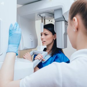
Dental Cone Beam CT
Dental cone beam computed tomography (CT) is a special type of x-ray equipment used when regular dental or facial x-rays are not sufficient. We use this technology to produce three-dimensional (3-D) images of your teeth, soft tissues, nerve pathways and bone in a single scan.
This procedure requires little to no special preparation. Please let us know if there’s a possibility you are pregnant.
Below are some commonly asked questions to guide our patients!
What is Dental Cone Beam CT?
Dental cone beam computed tomography (CT) is a special type of x-ray machine used in situations where regular dental or facial x-rays are not sufficient. The CT scanner uses a special type of technology to create 3D images of dental structures, soft tissues, nerve paths and bone in the craniofacial region in one scan. We are able to provide more precise treatment planning with images from cone beam CT scans.
The Cone Beam CT Difference
Cone beam CT is not the same as a conventional CT. Dental cone beam CT can produce images that are similar to those by conventional CT imaging.
With cone beam CT, an x-ray beam in the shape of a cone is moved around the patient to produce a large number of images, also known as views. CT scans and cone beam CT both produce high-quality images.
Dental cone beam CT was developed as a means of producing similar types of images but with a much smaller and less expensive machine that could be placed in an outpatient office.
Cone beam CT provides detailed images of the bone and is performed to evaluate diseases of the jaw, dentition, bony structures of the face, nasal cavity and sinuses. It does not provide the full diagnostic information available with conventional CT. Specifically, in evaluation of soft tissue structures such as muscles, lymph nodes, glands and nerves. However, cone beam CT has the advantage of lower radiation exposure compared to conventional CT.
What are some common uses of the procedure?
Dental cone beam CT is commonly used for treatment planning of orthodontic issues. It is also useful for more complex cases that involve:
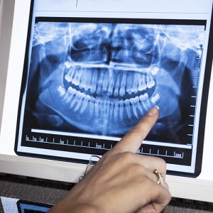
Digital X-rays offer a huge advantage in early detection and preventive services. Advanced image manipulation and analysis, allows your doctor to detect early stages of gum disease. This is done through recognition of bone level changes, or by evaluating the results against a previous treatment.
Enhanced with AI Technology
We use advanced AI-powered diagnostic tools alongside digital X-rays to help identify issues earlier and with greater accuracy. The AI software supports our doctors by highlighting subtle changes and patterns that may not be immediately visible to the human eye—enhancing diagnostic confidence and improving patient outcomes.
Benefits of Digital X-Rays
Benefits of digital x-rays compared to traditional dental X-rays include the following:
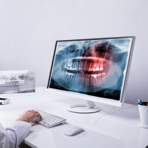
Electronic Dental Records are an integrated part of our practice management system. Our paperless system delivers electronic charting, digital imaging and enhanced case presentation where it is needed most…at chair side.
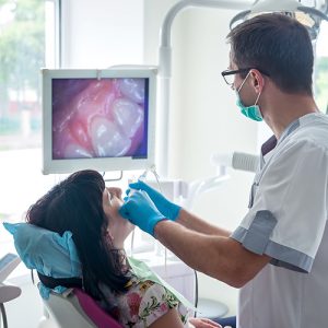
An intraoral camera is an indispensable diagnostic and educational tool. The tiny video camera moves around inside of your mouth and generates a tooth by tooth video exam of your teeth. Using an intraoral camera allows patients to better understand their dental needs by seeing what the dentist sees and then having it explained.
How Does It Work?
The intraoral camera resembles a oversize pen. The devices takes images of each tooth. While simultaneously viewing a monitor, the dentist inserts the camera into a patient’s mouth and gently shifts it about so that images can be taken from a variety of angles.

What is the T-Scan?
The T-Scan is a revolutionary device that measures the force and pressure between your upper and lower teeth. By measuring the force of your bite, we are able to spot occlusion problems that require treatment. In the past, the only way for us to identify issues with bite was with a small piece of blue contact paper. The T-Scan allows us to measure your occlusion to identify areas where the teeth are sitting incorrectly, causing unnecessary pressure and problems.
Why is the T-Scan needed?
When you have an occlusion problem, you may experience a wide range of different symptoms. For some patients, they grind their teeth regularly because their smile isn’t in alignment. For others, they might have tooth pain, headaches and even TMJ-related discomfort because of how they bite down. The T-Scan doesn’t allow for any guesswork, as it gives us a clear view of where your occlusion issues are located.
Who is a candidate for the T-Scan?
The T-Scan is painless and quick, so it’s ideal for most of our patients. Detection of an occlusion problem using the T-Scan can help when it comes to receiving quick, adequate treatment. The scan itself takes just minutes in our office and provides us with an in-depth view of your dental health. If you are experiencing occlusion problems or we suspect that there is an issue, we will recommend having a T-Scan done.
What happens during the T-Scan
We will sit you comfortably in one of our operatories. We then have you bite down on a thin device that electronically measures the amount of force between your upper and lower teeth. We use this data to help provide you with proper treatment of an occlusion problem. We will provide you with the results from the T-Scan and discuss ways to help improve your dental health. The T-Scan has been used on thousands of patients with wonderful success, and it is a great tool in eliminating issues with TMJ dysfunction, bruxism, tooth pain, dental damage and recurrent headaches.
If you’d like to come into our office to benefit from the T-Scan, call us today so that one of our helpful team members can further assist you.
What is Joint Vibration Analysis?
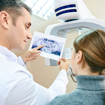
The temporomandibular joint is the area where the lower jaw attaches to the skull. When this joint becomes disorders, it can cause it to grind and scrape against itself. This causes vibrations that can be found through joint vibration analysis. By using this type of testing, we are able to determine what type of TMJ disorder you’re experiencing and the best way to go about treating it.
Why is Joint Vibration Analysis needed?
For some patients, their TMJ is painful, tender, swollen and stiff. This can have a serious impact on your quality of life. A regular examination as well as x-rays often aren’t enough to truly understand the problem at hand. For this reason, we recommend joint vibration analysis so that we are able to identify the vibrations being caused by joint friction.
Who is a candidate for Joint Vibration Analysis?
Most patients who suffer from TMJ disorder are good candidates for joint vibration analysis. The analysis is done right in our office and can be beneficial in that it takes just minutes. We are then able to identify where and why your joint is experiencing dysfunction and the best way to go about treating it. The analysis is easy, quick and provides us with highly detailed results.
What happens during Joint Vibration Analysis?
Joint vibration is quick, easy and painless. We first sit you down in our office and use a small handheld machine that is placed against the TMJ. We then use this machine to measure the amount of vibrations being causes when opening and closing your mouth. The amount of vibrations can give us a clear view of the problem you’re experiencing. Once we utilize this analysis, we can determine the best course of treatment for your TMJ dysfunction. Treatment is more effective and can benefit your overall quality of life by improving TMJ function each day.
If you would like to learn more about joint vibration analysis, call us today so that we can get you in for a convenient consultation appointment.
What is 3D Printing?
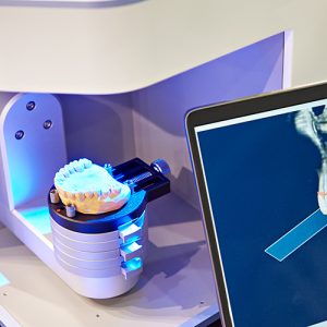
3D printing is a revolutionary way for us to better treat our patients. We use this printing to help create surgical guides so that treatment planning is easier and more effective. The printing takes digital or impression renderings of your teeth and mouth to create a model that we can actually hold and work with when planning your treatment. The process of making a 3D model of your teeth is easy, quick and highly effective when it comes to your dental care.
Why is 3D Printing needed?
The reason we may recommend 3D printing in our office is because you’re going to be having oral surgery done. For instance, having a 3D model of your teeth and mouth can help us in determining the best placement for implants or when performing a bone grafting. The rendering is realistic and detailed, so we are able to pinpoint the different ways to complete the surgical procedure.
Who is a candidate for 3D Printing?
Taking a 3D impression of your teeth is easy and quick. Most of our patients can benefit from 3D printing just because of how beneficial it is to their overall future dental health. Both children and adults alike can have 3D renderings done of their teeth. We will be able to recommend when and if this printing is needed during the course of your treatment.
What can be expected with 3D Printing?
The first step is to take impressions of your teeth and mouth. This is typically done digitally so that we have a clear view of your oral health on our computer screens. We then use a 3D printing machine to create a physical model of these impressions. We then use this model to help in treatment planning for surgical procedures. We will help to show you what to expect during oral surgery using this model as well. 3D printing is highly useful when it comes to improving the patient experience.
If you’d like to learn more about 3D printing and how it works, call our office today and one of our trained staff members will be more than happy to help assist you.